


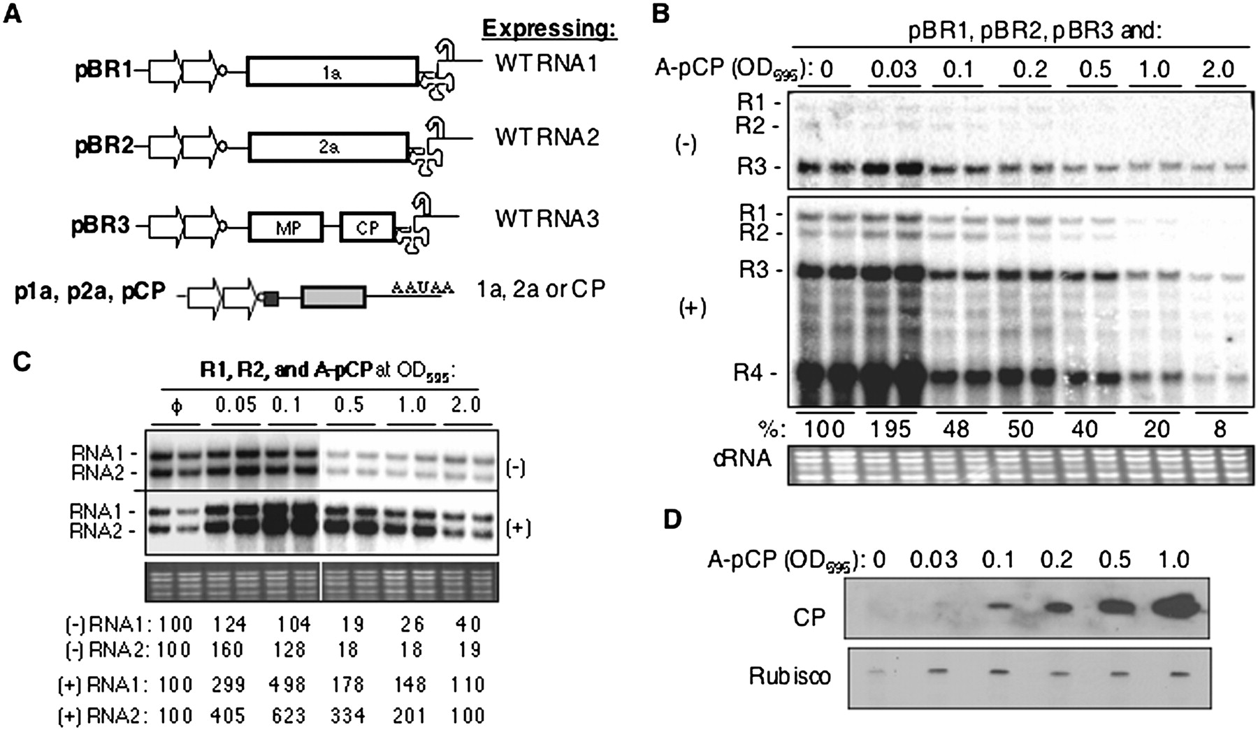
The BMV capsid protein (CP) can regulate BMV RNA accumulation. (A) Schematic of the constructs used in this study. The relevant portions of the T-DNA plasmids are shown with the names to the left. The two arrows represent the tandem CaMV 35S promoters used to drive transcription. The named of protein(s) encoded by the RNAs are shown inside rectangles. The 3′ UTR is represented by a cloverleaf. The curved arrow represents a cis-acting ribozyme that will generate the bona fide 3′ terminus of the BMV RNAs. The cDNAs used for transient expression of BMV proteins 1a, 2a, and CP contain a translational enhancer in the 5′ UTR upstream from the protein-coding sequence (small black square) and a polyA processing signal (AAUAA) 3′ of the protein-coding sequence. (B) Effects of transient BMV CP expression on BMV RNA accumulation. Leaves of N. benthamiana plants were infiltrated with a mixture of Agrobacterium cultures expressing BMV CP with a mixture of cultures that expressed three genomic RNAs (at OD595 of 0.1). Total RNA was isolated at 48 hpi, and minus- and plus-strand viral RNAs were detected by Northern blot using strand specific probes as described by Gopinath et al. (2005). The agarose gel image of ethidium bromide-stained cellular RNA (cRNA) served as an RNA loading control. Quantification of the minus- and plus-strand RNAs shown as percentages of the wild-type RNAs in B. (C) Effects of the CP on BMV RNA1 and RNA2 replication. N. benthamiana leaves were infiltrated with Agrobacterium cultures expression BMV RNA1 and RNA2 (at an OD595 of 0.1). Total RNAs were isolated and analyzed as described in B. (D) Western blot of CP expression level in N. benthamiana plants infiltrated with different concentrations of Agrobacterium expressing BMV CP. The primary antibody was a rabbit polyclonal antibody purchased from the ATCC. A Coomassie blue-stained small subunit of Rubisco was used as a protein loading control.




