


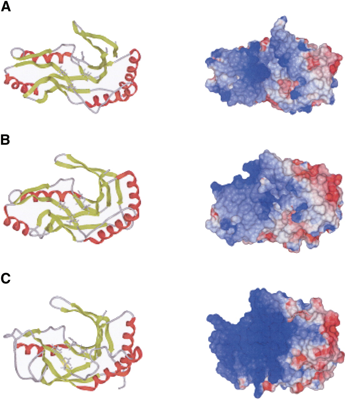
FIGURE 8.
Comparison of tertiary structure, (left) HXTX motif, and (right) electrostatic surface potential of PF0027 and two structurally related proteins. (A) PF0027 (PDB entry 2FYH), (B) PF0027-related protein from Pyrococcus horikoshii (PDB entry 1VDX), and (C) PF0027-related protein from Thermus thermophilus (PDB entry 1IUH). (Red) The α-helices and (light green) β-strands. The histidine and threonine residues in the HXTX motif are shown in stick model format. Images were produced by ICM Pro (Molsoft Inc.). Another structure for the PF0027-related protein from P. horikoshii (PDB entry 1VGJ) is essentially the same as the 1VDX structure shown.




