


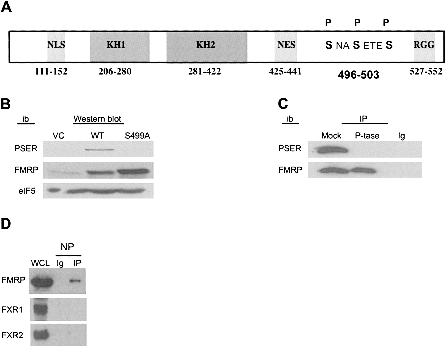
PSER antibody is specific for phosphorylated FMRP. (A) Schematic representation of the FMRP. NLS-nuclear localization sequence, NES-nuclear export sequence, phospho-serines 496, 499, and 503 (adapted from data in Ceman et al. 2003). (B) Western blot of whole-cell lysates from VC, WT, and S499A FMRP expressing cells were immunoblotted (ib) with the indicated antibody: PSER, 1a (anti-FMRP 1a-1C3) (Devys et al. 1993), or eIF5. (C) Anti-Flag immunoprecipitations (IP) of Flag-FMRP expressing L-M[TK-] cells were split and mock-treated or phosphatase (P-tase)-treated (Ceman et al. 2003) before probing with PSER or 1a (anti-FMRP). Ig-immunoprecipitating antibody alone. (D) Flag-FMRP expressing L-M[TK-] cells immunoprecipitated with NP antibody and probed with 1a (anti-FMRP), FXR1, or FXR2 antibodies. WCL-L-M[TK-] whole-cell lysate. Ig-NP indicates antibody alone.




