


Cover image
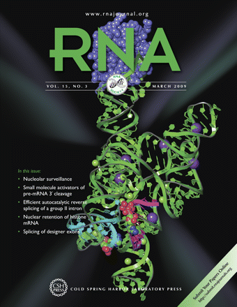
On the cover
Cover Illustration: Crystal structure of the group I intron P9 complexed with spliced exons (deoxy post-25 construct). (Protein Data Bank code: 3bo4; Lipchock, S.V. and Strobel, S.A. 2008. A relaxed active site after exon ligation by the group I intron. Proc. Natl. Acad. Sci. 105: 5699–5704.) Image details: RNA binding domain of the U1A protein (aids crystallization): space–filling representation—blue; group I intron P1: ribbon–plate representation, green; ligated exons: ribbon-plate representation—cyan; U1 small nuclear ribonucleoprotein A RNA: ribbon—plate representation—yellow. The image was generated with PyMOL (DeLano Scientific LLC). Cover image provided by the Jena Library of Biological Macromolecules —JenaLib (www.fli-leibniz.de/IMAGE.html).


Molecular Cloning
The New Edition
Buy Now and Save 30%
Paperback only
(limited time offer)

|
Molecular Cloning: A Laboratory Manual (Fourth Edition) |