


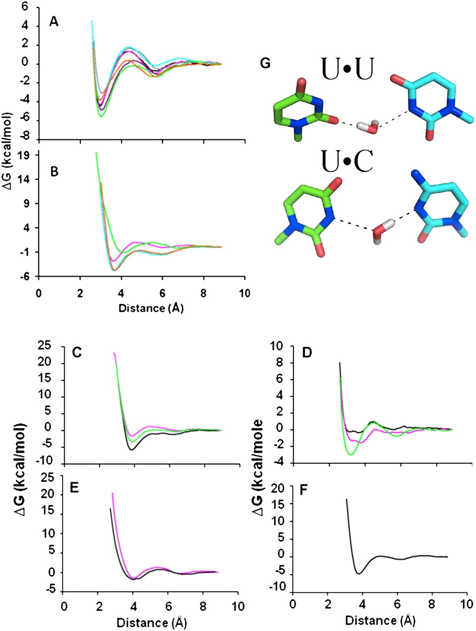
Free energy change as a function of hydrogen bond distance for U•A, U•G and modified U base. (A) Free energy change as a function of hydrogen bond distance of the Watson–Crick and the modified U•A base pairs. Distance and energy relationships for the base-pairing of the unmodified U•A ribonucleoside-5′-monophosphates (black) and the modified uridines with A are very comparable: s2U•A (green), mnm5U•A (cyan), mnm5s2U•A (gray), mcm5s2U•A (purple), cmo5U•A (pink), Ψ•A (orange). It should be noted that the curves for cmo5U•A (pink) and U•A (black) are almost identical and obscure the lines. (B) Free energy change as a function of hydrogen bond distance of the modified wobble U•G base pairs. U•G base pairs include the modified Ψ•G (orange), cmo5U•G (pink), mnm5U•G (cyan), and s2U•G (green). (C) U•U mismatch base pairs including modified cmo5U•U (pink), s2U•U (green), and unmodified U•U (black). (D) U•C base pairs of modified cmo5U•C (pink), s2U•C (green), and unmodified U•C (black). (E) Free energy diagram of the modified mnm5s2U•G (black) and mcm5s2U•G (pink) base-pairings. (F) Free energy change as a function of hydrogen bond distance for the Ψ•U base pair. (G) Stabilization of pyrimidine•pyrimidine base-pairings by a water bridge. A water molecule bridges the pyrimidine•pyrimidine base pairs of U•U and U•C. The nonprotonated nitrogens are hydrogen bonded to the exchangeable hydrogens on the water, thus stabilizing the interaction of the bases. The resulting base-pair exhibits a ribose C1′–C1′ distance comparable to that of A-form RNA helices. Stick models have oxygen in red, nitrogen in blue, and hydrogen of the water in gray.




