


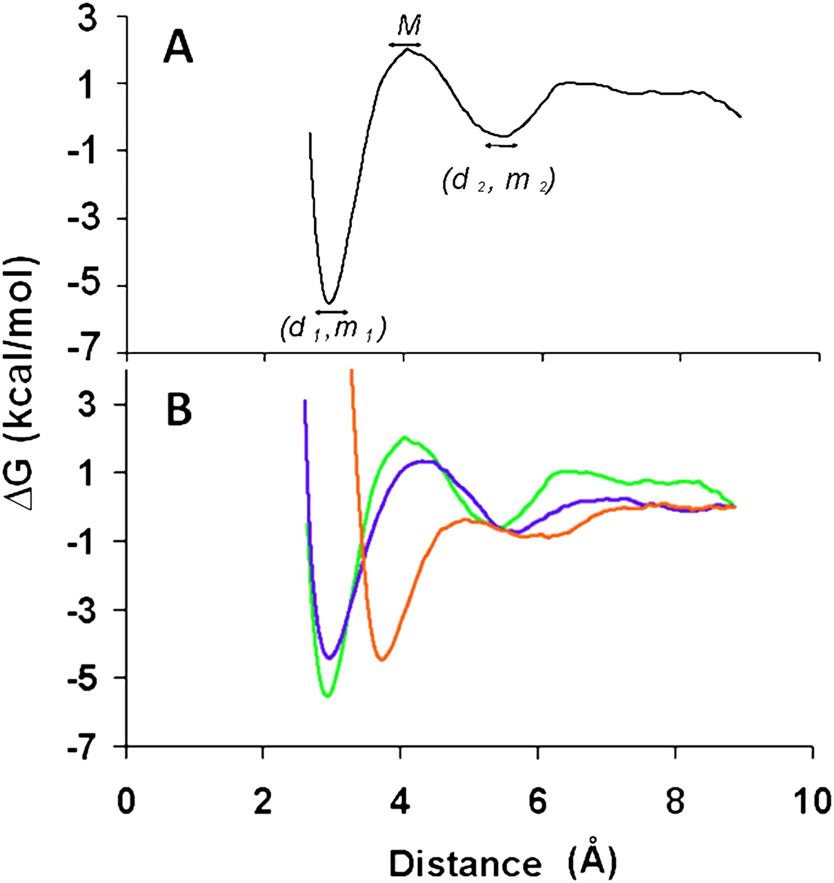
Free energy variation as a function of hydrogen bond distance. (A) Free energy variation as a function of hydrogen bond distance for the molecular dynamics simulation (MDS) of a hypothetical base-pairing by ribonucleoside-5′-monophosphates. The global minimum of the base-pair energy, the local minimum of that energy, and energy maximum are displayed and are denoted as m1, m2, and M (kcal/mol), respectively. The hydrogen bond distances between the bases for the imino proton hydrogen bond correspond to d1 and d2 (Å). (B) Free energy for the Watson–Crick base-pairings of the ribonucleoside-5′-monophosphates U•A and C•G and for the wobble U•G base pair. The MDS of base-pairing was conducted in an aqueous environment under neutral conditions with Na+ as the counter ion to the ribonucleoside-5′-monophosphate. The Watson–Crick base pairs C•G (green), U•A (blue) exhibited comparable energy/distance relationships with the C•G pair being the more stable of the two. The wobble pair U•G (orange) exhibited a different profile, with a longer imino hydrogen bond distance. All three base-pairings exhibited a secondary interaction resulting in an intermediate stability (m2) and longer distance (d2) between bases bridged by one or two H2O molecules.




