


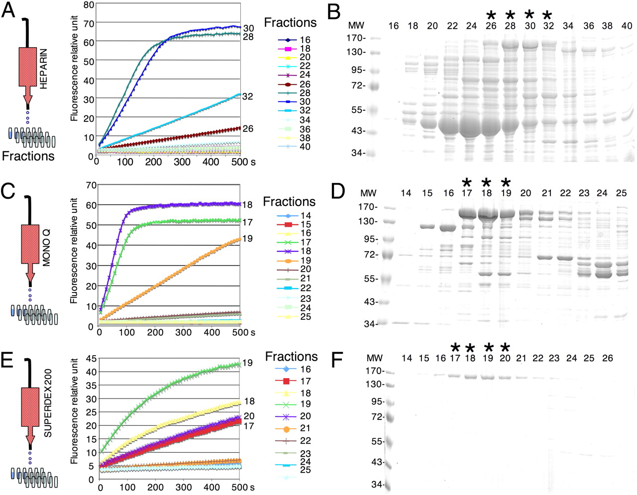
Purification of a 5′–3′ exoribonuclease, Xrn1 from S. cerevisiae. The protocol used for this purification is identical to a previously published protocol (Pellegrini et al. 2008) using radiolabeled RNA to test for the Xrn1 activity. Samples from successive elutions (see Materials and Methods), Heparin (A), Mono Q (C), and Superdex 200 (E), were diluted 100-fold and assayed in the presence of the fluorogenic RNA1/DNA1 duplex. Only samples of interest are shown. After the purification, samples were analyzed by Coomassie blue-stained SDS-PAGE (heparin [B]), Mono Q [D], and Superdex 200 [F] showing the successive steps of Xrn1 purification (band at 175 kDa). Equal volumes of dilutions of the different fractions were assayed independently of total protein concentrations. For example, the fraction 19 (C) has an apparently lower activity than fractions 17 and 18, but this fraction is less concentrated in Xrn1 protein (B). (MW) Protein size standard in kilodaltons. Lanes noted with an asterisk (*) correspond to fractions with peaks of Xrn1 activity.




