


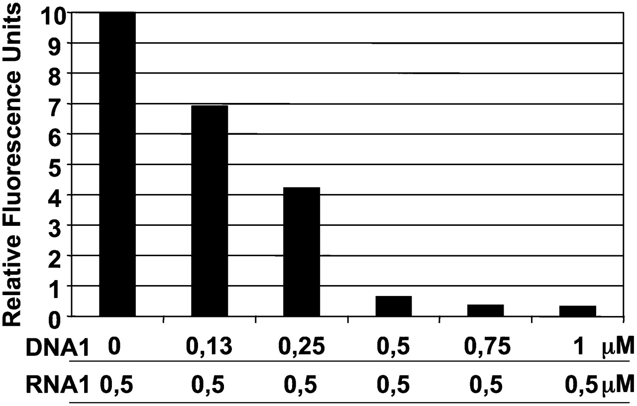
FIGURE 2.
Fluorescence quenching of FAM by TAMRA. The following assays were monitored on a Rotor Gene apparatus at 30°C, set up to detect FAM fluorescence. A concentration of 0.5 μM of RNA1 (FAM-linked) was incubated with various concentrations of DNA1 (TAMRA-linked). At 30°C, quenching of FAM fluorescence confirms that the duplex forms instantly (the duplex was designed with a Tm of 34°C). A stoichiometry of two molecules of TAMRA for one molecule of FAM leads to a 30-fold reduction in the fluorescence emitted by the FAM-linked RNA. Reproducible results were obtained with various RNA1 concentrations (ratio 1/2 for FAM/TAMRA).




