


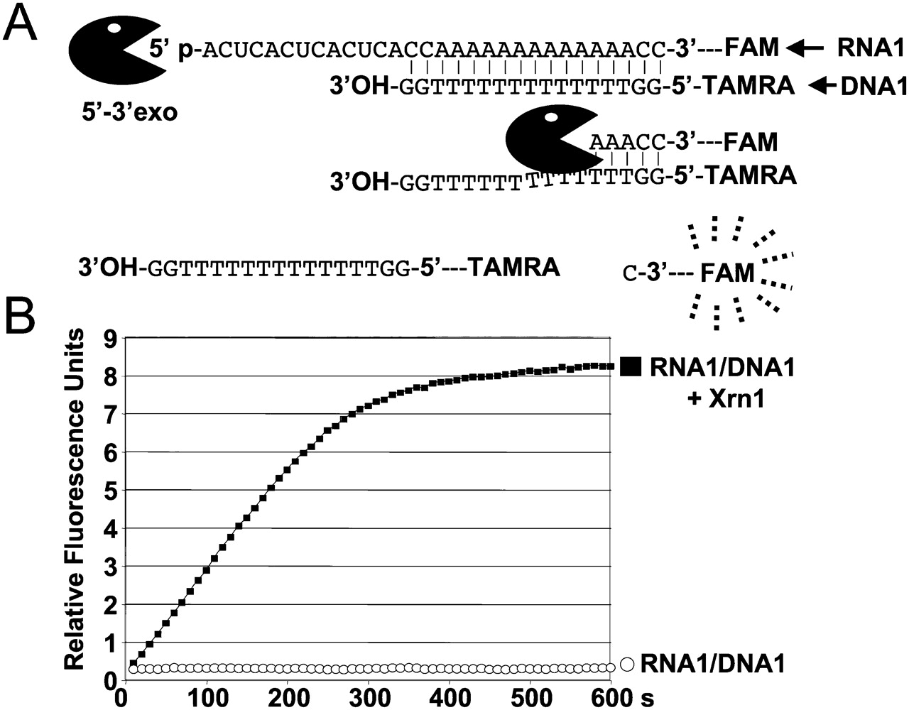
FIGURE 1.
Real-time fluorescence-based analysis of RNA hydrolysis by Xrn1 exoribonuclease. (A) The formation of the RNA–DNA duplex brings the TAMRA into the vicinity of FAM to quench FAM fluorescence emission (see Fig. 2). Digestion of RNA1 releases the FAM from the oligonucleotide and permits FAM fluorescence to be detected at 520 nm. (B) In the presence of 50 nM Xrn1 exoribonuclease and 500 nM of RNA1, an immediate generation of FAM fluorescence signal can be monitored in real time.




