


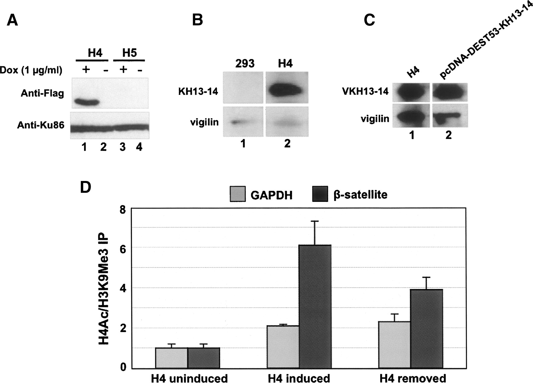
(A) Western blotting with anti-Flag antibody on lysates from expanded hygromycin resistant clones from stable cell lines expressing flag-KH13–14. Ku86 was used as loading control. Clone H4 expresses flag-KH13–14, while clone H5 does not. (B) Expression of flag-KH13–14 in the H4 cell line was confirmed by Western blotting with anti-vigilin antibody that recognizes the C-terminal portion of vigilin. Expression of endogenous vigilin was used as loading control. HEK293 cells (“293”) were used as control. (C) Lysates from H4 cells or from HEK293 cells transiently transfected with pcDNA-DEST53-KH13–14 were analyzed by Western blotting with anti-vigilin antibody. Expression of endogenous vigilin was used as loading control. (D) Chromatin changes in cells with inducible KH13–14. Equal amounts of lysates from uninduced H4 cells, induced H4 cells, or induced H4 cells removed from doxycycline treatment were used for ChIP assays with antibodies against trimethylated H3K9 and acetylated H4, followed by PCR with primers specific for β-satellite or GAPDH sequences. Chromatin status is presented as the ratio of fold enrichment of PCR signals from H4Ac IP to that from H3K9Me3 IP. The ratios obtained from induced stable cell ChIP analyses were normalized to those from uninduced stable cells.




