


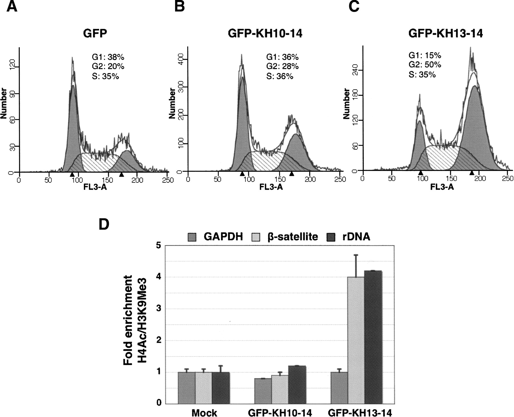
(A–C) The KH13–14 fragment exhibits dominant negative effects on chromosome segregation. HEK293 cells were transfected with plasmids expressing GFP, GFP-KH10–14, or GFP-KH13–14. Seventy-two hours after transfection, GFP-positive cells were collected and analyzed by FACS. Compared to control cells transfected with GFP alone (A) or GFP-KH10–14 (B), cells transfected with GFP-KH13–14 (C) are mostly arrested at the G2/M phase, with an increased average DNA content indicated by the intensity of PI staining of DNA (black arrows). The data shown are representative results from three independent experiments. (D) Antibodies against trimethylated H3K9 or acetylated H4 were used for chromatin immunoprecipitations with sheared chromatin from untransfected HEK293 cells or cells transfected with GFP or GFP-tagged vigilin mutants. Purified DNAs were used for real time PCR with primers specific for pericentromeric β-satellite, rDNA, or GAPDH sequences. The chromatin status of these regions is expressed as the ratio of PCR signal obtained for H4Ac (euchromatin marker) IP to that obtained for H3K9Me3 (heterochromatin marker) IP. The ratios from transfected cells were normalized to those from untransfected cells. The results represent mean ± SD signal of PCRs performed in triplicate.




