


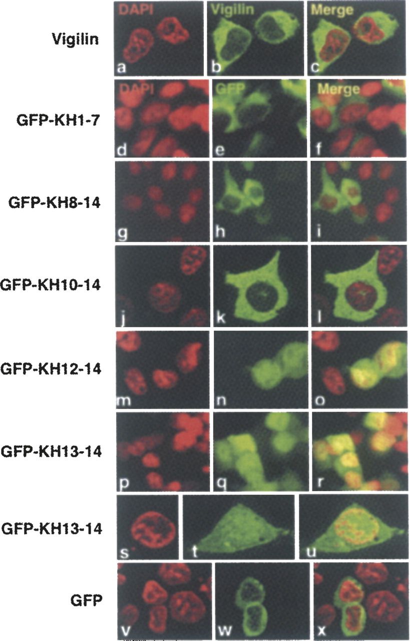
FIGURE 3.
Immunolocalization of vigilin and vigilin fragments. (a–c) Immunolocalization with a polyclonal anti-vigilin antibody. DAPI is pseudocolored in red. For panels d–x the indicated GFP-tagged constructs were transfected into HEK293 cells and GFP expression monitored and localized by fluorescence. Note that GFP-KH1–7, GFP-KH8–14, and GFP-KH10–14 are mostly in the cytoplasm, while GFP-KH12–14 and GFP-KH13–14 are equally distributed between the cytoplasm and nucleus.




