


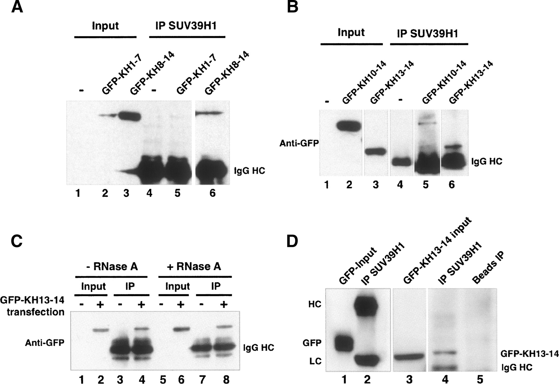
Mapping the SUV39H1 binding domain of vigilin. (A) HEK293 cells were transfected with GFP-tagged KH1–7 or KH8–14, IP was carried out with anti-SUV39H1 antibody as in Figure 1, and Western blotting was with anti-GFP antibody. (B) Cells were transfected with vigilin KH10–14 or KH13–14 followed by IP and Western blotting as in A. (C) The interaction of SUV39H1 with the vigilin KH13–14 region is independent of RNA. Cells were transfected with vigilin GFP-KH13–14. The same experiments were carried out as in B, except that cell lysates were treated with 200 μg/mL RNase A at 4°C for 2 h before immunoprecipitation. (D) SUV39H1 specifically associates with GFP-tagged vigiin KH13–14 (lane 4) but not GFP alone (lane 2) in vivo. Beads alone cannot pull down vigilin KH13–14 (lane 5). Cells were transfected with plasmid expressing GFP or GFP-KH13–14 followed by IP with anti SUV39H1 antibody and Western blotting with anti-GFP antibody.




