


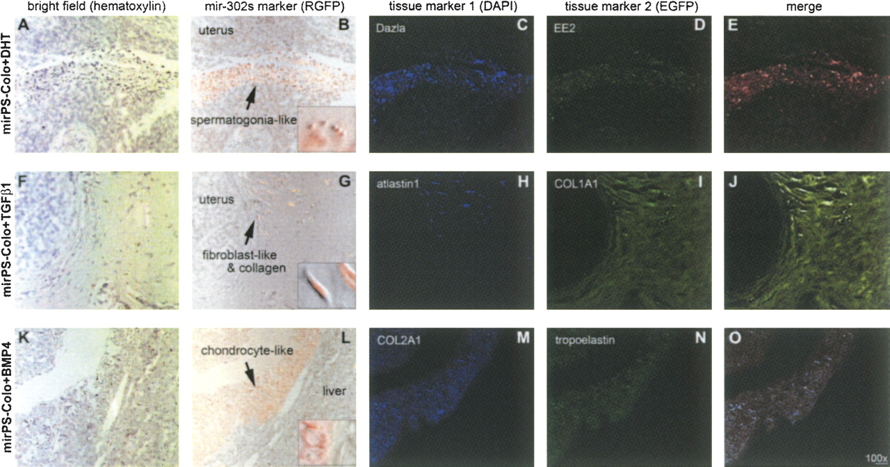
Pluripotency of mirPS cells. Treatments of DHT, TGF-β1 and BMP4, respectively from top to bottom, induced the mirPS-Colo cell differentiation into spermatogonia-like (A–E), fibroblast-like (F–J), and chondrocyte-like (K–O) primordial cells, in immunocompromised mice ex vivo. The use of immunocompromised nude mice was to provide an in vivo environment mimicking transplantation therapy. Microscopic photographs shown from left to right indicated hematoxylin staining with differential interference contrast (A, F, K), bright field labeled with transgenic mir-302 marker RGFP (red) (B, G, L), immunostaining of the first tissue marker labeled with 4,6-diamidino-2-phenylindole (blue DAPI) (C, H, M), immunostaining of the second tissue marker labeled with fluorescein (green EGFP) (D, I, N), and merge of all three fluorescent markers (E, J, O). Small windows in the RGFP–bright fields showed the morphologies of differentiated mirPS cells at high magnification (600×).




