


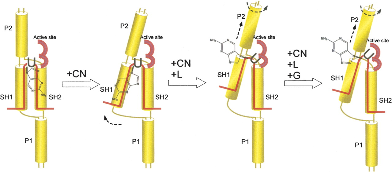
FIGURE 6.
Distinct substrate RNA conformations in partially and fully docked complexes are revealed by fluorescence studies. A cartoon representation of substrate conformations accounting for the observed fluorescence intensities. “U” denotes the target uridine and the fused rings represent 2-AP. Yellow cylinders represent the four helices formed by the guide–substrate RNA complex.




