


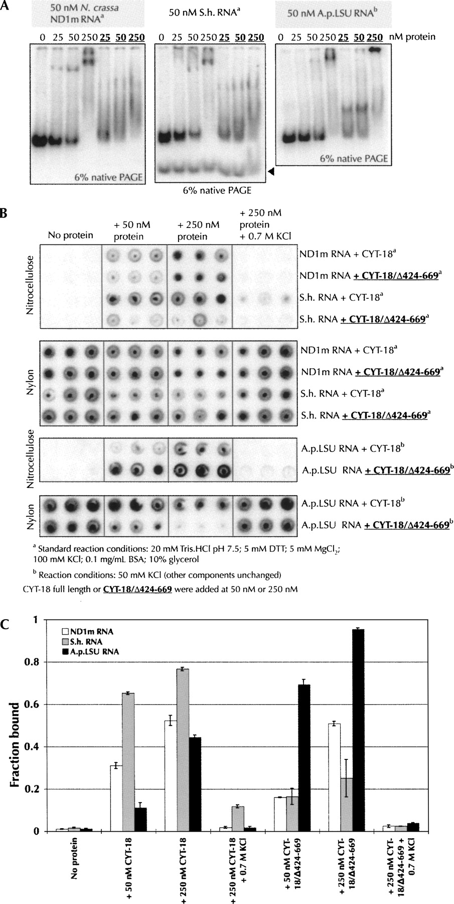
Binding assays of the full-length and C-terminally truncated CYT-18 proteins to the ND1m, S.h., and A.p.LSU introns. (A) Gel shift assays with 50 nM RNA and increasing concentrations (0, 25 nM, 50 nM, and 250 nM) of CYT-18 full length or truncated (bold underlined). In this and subsequent panels, CYT-18 binding and protein-facilitation assays were carried out in standard conditions for the ND1m and S.h. introns (20 mM Tris-HCl at pH 7.5, 5 mM DTT, 5 mM MgCl2, 100 mM KCl, 0.1 mg/mL BSA, 10% glycerol) and at 50 mM KCl for the A.p.LSU intron (other components unchanged). The black arrow indicates the linearized S.h. intron. (B) Filter binding assays upon addition of increasing concentrations (0, 50 nM, and 250 nM) of CYT-18 full length or tuncated (bold underlined). Experiments were performed in triplicate. (C) Quantitation of the filter binding experiments shown in panel B. The error bars indicate the standard deviation calculated from the triplicate experiments shown in B.




