


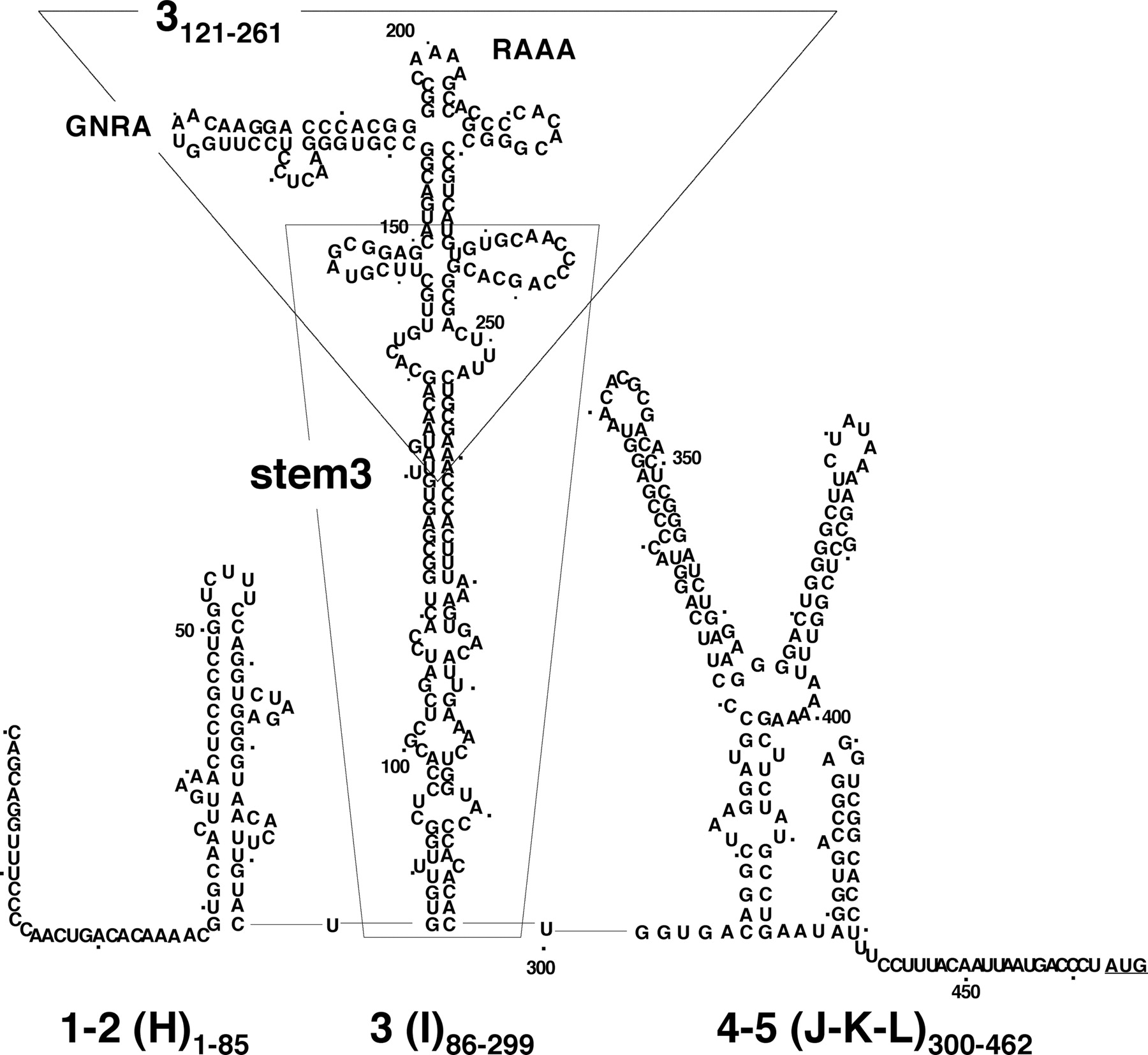
FIGURE 2.
Secondary structure of FMDV IRES. RNA structure is depicted according to RNA probing analysis (Fernandez-Miragall et al. 2006). IRES domains are numbered 1–5 (or H–L); the nucleotides corresponding to the IRES transcripts used in this study are indicated as subscripts; a dot is used to mark positions every 10 nt. The conserved GNRA and RAAA motifs in domain 3 are highlighted. A broken line surrounds transcript 3121–261. A thin line surrounds transcript stem3, encompassing residues 85–299 with 151–227 deleted.




