


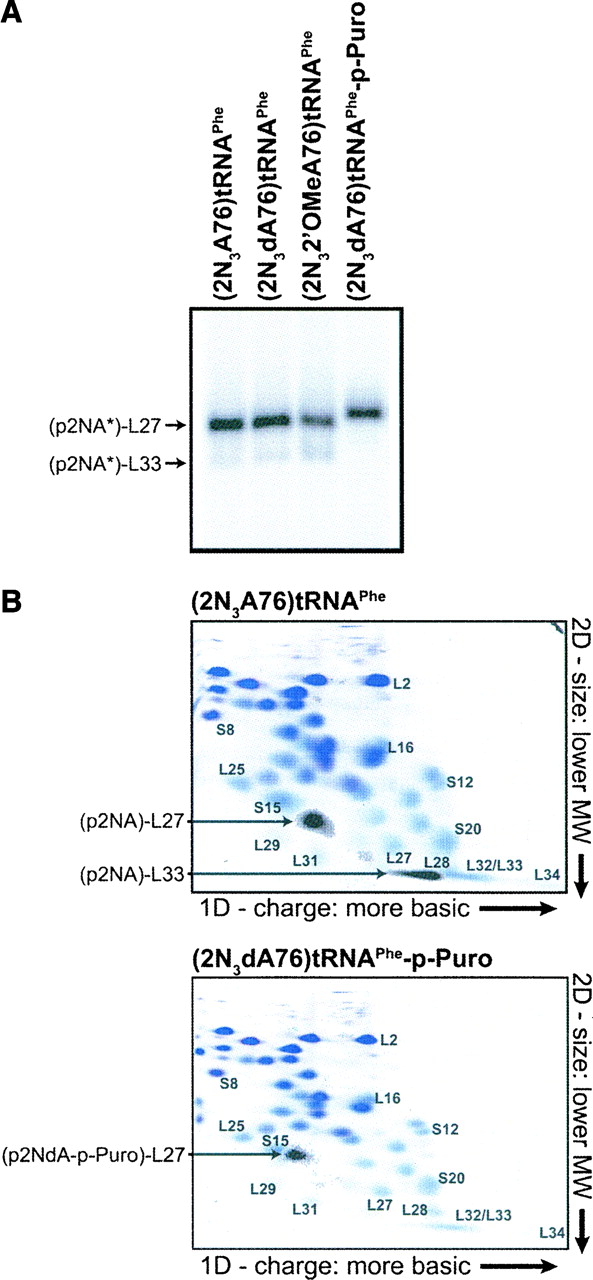
Identification of ribosomal proteins cross-linked by each photoreactive ligand. 50S-subunit components were solubilized in 6 M urea and incubated with ribonuclease S1, which entirely digested the RNA, leaving a radioactively labeled 5′-phosphate and its modified A76 nucleotide covalently attached to a ribosomal protein. The digested material was resolved by (A) 10% SDS-PAGE and by (B) two-dimensional PAGE, and then visualized by autoradiography. In panel A the labeled bands contain different derivatives of A76, collectively designated as A*. In panel B, the samples were spiked with nonradioactive proteins that were acid-extracted from purified 70S ribosomes to provide reference markers. The proteins in the two-dimensional (2D) gel were stained with bromophenol blue and the image was overlaid with the autoradiogram of the same gel. The positions of (p2NA)-L27, (p2NA)-L33, and (2NdA-p-Puro)-L27 are displaced from those of L27 and L33 owing to the presence of the negatively charged nucleotide moieties (Maguire et al. 2005).




