


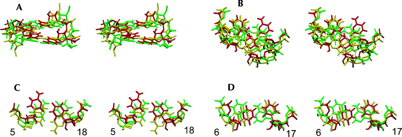
FIGURE 7.
Overlays of the lowest-energy structures of the poliovirus Y domain (Lescrinier et al. 2003) and variants (this work), superimposed on the central seven base pairs, i.e., residues 2–8 and 15–21. The poliovirus 1 structure (Lescrinier et al. 2003) is colored red, the Ycu variant green, and the Yuu variant yellow. (A) Stereo side view into the major groove of the two central residue 5–18 and 6–17 base pairs. (B) Stereo top view down the helical axes. The residue 6–17 base pairs are on top. (C) Stereo top view of the residue 5–18 base pairs. (D) Stereo top view of the residue 6–17 base pairs.




