


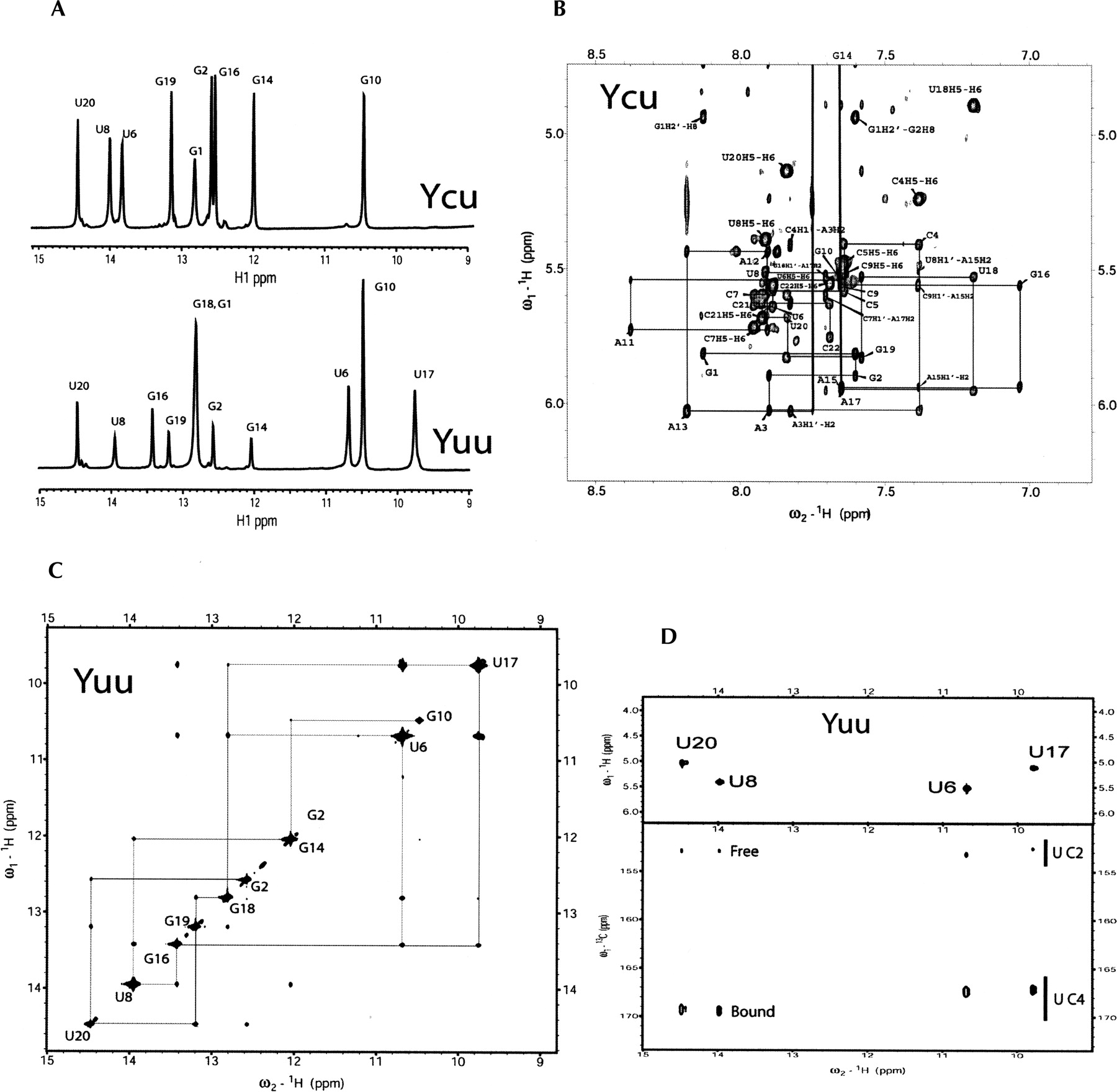
(A) One-dimensional 1H NMR spectra of the Yuu (lower trace) and Ycu (upper trace) RNA hairpins recorded at pH 6.0 at 600 MHz, showing the imino proton region. Assignments to residues are indicated. (B) Section of the 2D NOESY spectrum (300 msec mixing time) of the Ycu RNA hairpin recorded in D2O at pH 6.8 at 800 MHz, showing the anomeric to aromatic proton region. Sequential H1′-H8/H6 connectivities are indicated by lines. Intranucleotide H1′-H6/H8 NOEs are annotated according to residue type and number, e.g., G1. (C) Section of the 2D NOESY spectrum (300 msec mixing time) of the Yuu RNA hairpin recorded in 90% H2O/10% D2O at pH 6.0 at 800 MHz, showing the imino proton region. Assignments of the diagonal peaks are indicated by residue type and number. The sequential imino-to-imino proton walk is traced out. (D) Residue-specific imino proton assignment and chemical-shift dependence of the C2 and C4 chemical shifts on base pairing of the Yuu RNA hairpin. The spectra were recorded at 5°C at 600 MHz. The 2D back-and-forth H(N3C5)H5 spectrum on top shows the imino-H5 connectivities of the U-specific 13C/15N-labeled Yuu sample, used for assignnment of uridine imino protons. The 2D HNCO spectrum on the bottom was used to establish the hydrogen bonding patterns of the uridines based on the C2 and C4 chemical shifts.




