


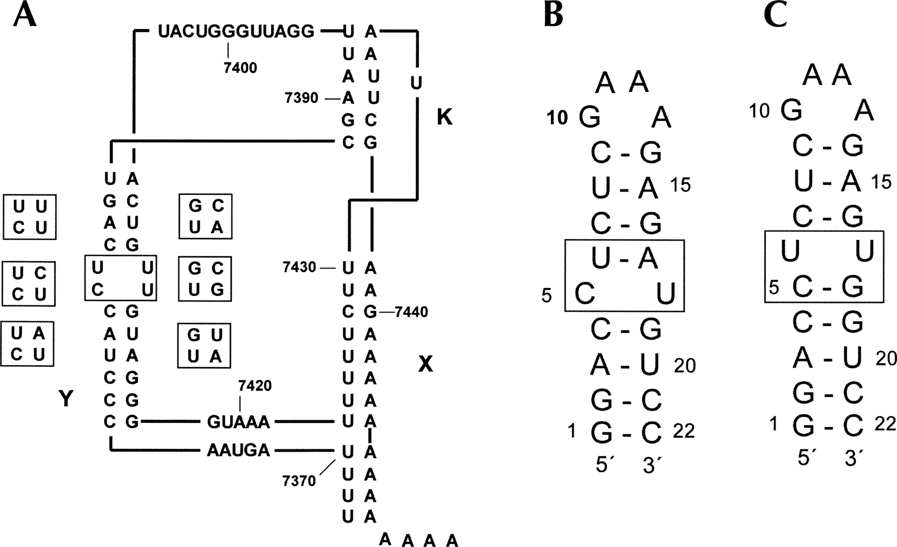
FIGURE 1.
(A) Schematic representation of the secondary structure of the 3′-UTR of poliovirus type 1. (B) RNA hairpin Ycu and (C) hairpin Yuu used for the NMR studies. Variation of the central two base pairs in the Y domain is shown on the left of the Y domain in A for the polio and polio-like coxsackie viruses, for the nonpolio-like coxsackie and ECHO viruses on the right.




