


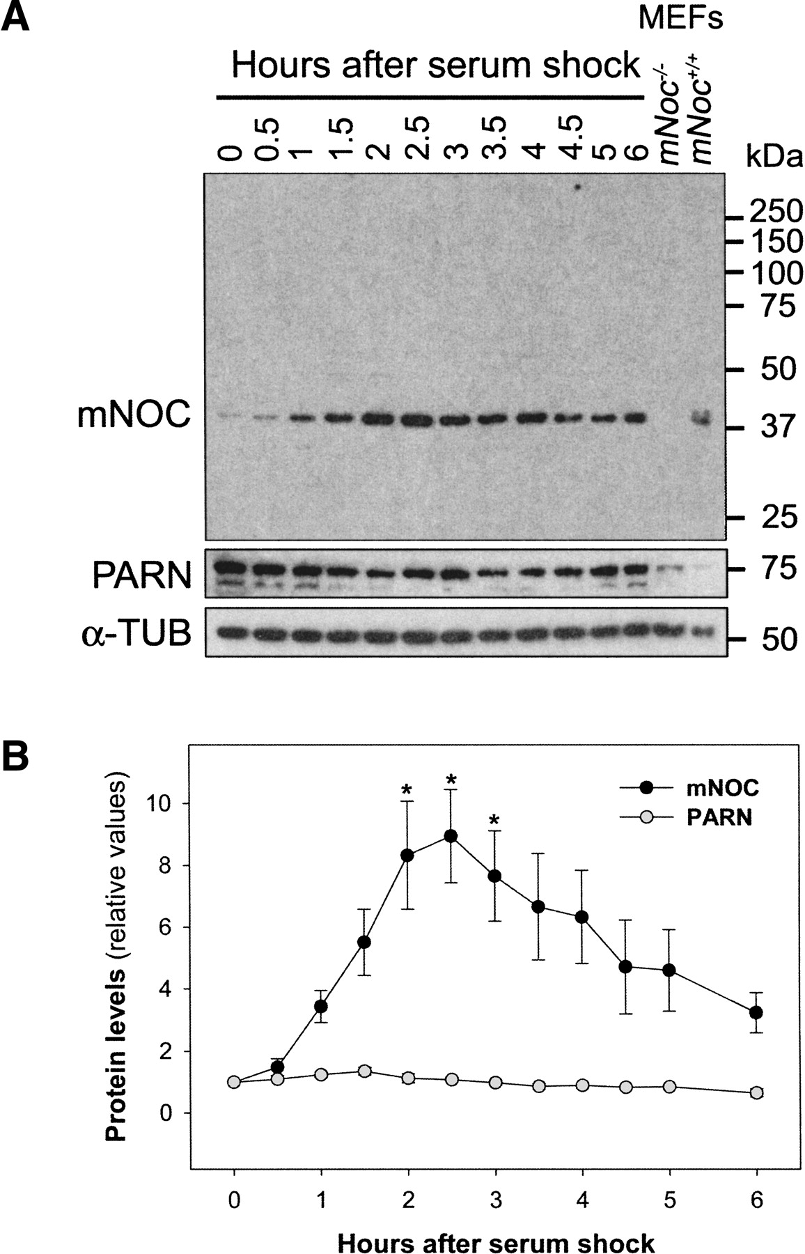
FIGURE 6.
mNOC protein induction by serum shock. (A) Western blot showing the temporal profile of the mNOC and PARN protein levels after a serum shock. Cells were treated as in Fig 5. The lanes containing lysates of mouse embryo fibroblasts (MEFs) obtained from mNoc+/+ and mNoc−/− mice serve as positive and negative controls. MEFs were serum stimulated for 2 h. In the lower panel, an immunoblot of the same membrane using α-tubulin antiserum as a loading control. (B) The results of three blots were quantified with the Scion Image software; the graph shows the mean ± SEM. (*) p < 0.05.




