


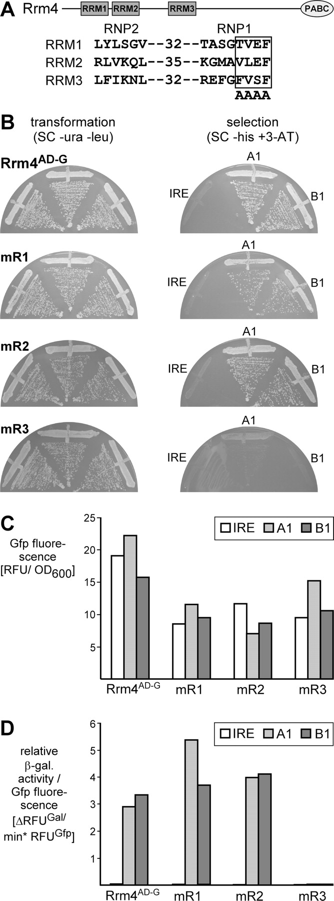
Identifying the aptamer interaction domain of Rrm4 in vivo. (A) Schematic representation of the Rrm4 domain architecture (top; RRM and C-terminal PABC domains are given as rectangles and oval, respectively) and the introduced point mutations (bottom). The boxed amino acids in the RNA contact region RNP1 were mutated to alanine. The numbers indicate the distance between RNP1 and RNP2. (B) HIS3 expression was monitored as growth on selection plates (SC −his+5 mM 3-AT). Strain L40-coat expressed different versions of Rrm4AD-G (Rrm4AD-G-mR1, Rrm4AD-G-mR2, and Rrm4AD-G-mR3 carry point mutations in RRM1, RRM2, and RRM3, respectively; labeled with mR1, mR2, and mR3 on the left). The respective hybrid RNA is indicated next to the various plate sectors (labeled on the right). (C) Expression of Rrm4AD-G versions (given at the bottom) was determined in strains expressing different hybrid RNAs (inlay) by quantifying Gfp fluorescence per OD600. (D) Relative β-galactosidase activity was determined using a fluorogenic substrate. To account for differences in protein expression, enzyme activity is given relative to Gfp fluorescence (ΔRFUGal per minute per RFUGfp). Different versions of Rrm4AD-G (bottom) were tested with various hybrid RNAs (inlay).




