


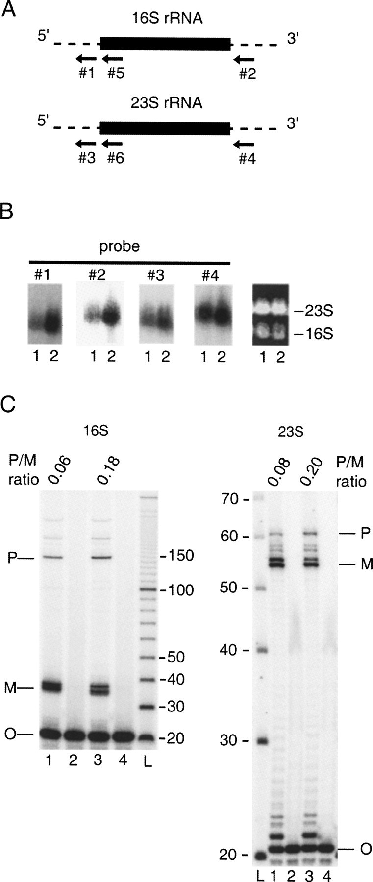
Ribosomal RNA processing defects in HMSdrp. (A) Schematic description of oligonucleotide probes used to assess rRNA processing by Northern blot analysis and primer extension. Probes #1–4 are partially or fully complementary to 16S or 23S rRNA precursor sequences (dashed lines) that are absent in mature rRNAs (thick lines). Probes #5–6 anneal 15–35 and 35–55 nt downstream from the mature 16S and 23S rRNA 5′ ends, respectively. (B) Northern blot analysis of RNA samples isolated from HMS174(DE3) (lane 1) and HMSdrp (lane 2) with probes #1–4 (four left panels). (Right panel) The agarose gel stained with ethidium bromide. The positions of 16S and 23S rRNAs are indicated. (C) Primer extension of total RNAs from HMS174(DE3) (lanes 1,2) and HMSdrp (lanes 3,4) using probes #5 and #6, specific for 16S and 23S rRNA, respectively. (Lanes 2,4) Control lanes where no reverse transcriptase was added. (Lane L) A 10-bp ladder. The positions of the oligonucleotide probes (O), and the reverse-transcription products derived from the mature (M) and precursor (P) rRNAs are noted. The ratios of the amounts of precursor to mature rRNAs are indicated above the gels.




