


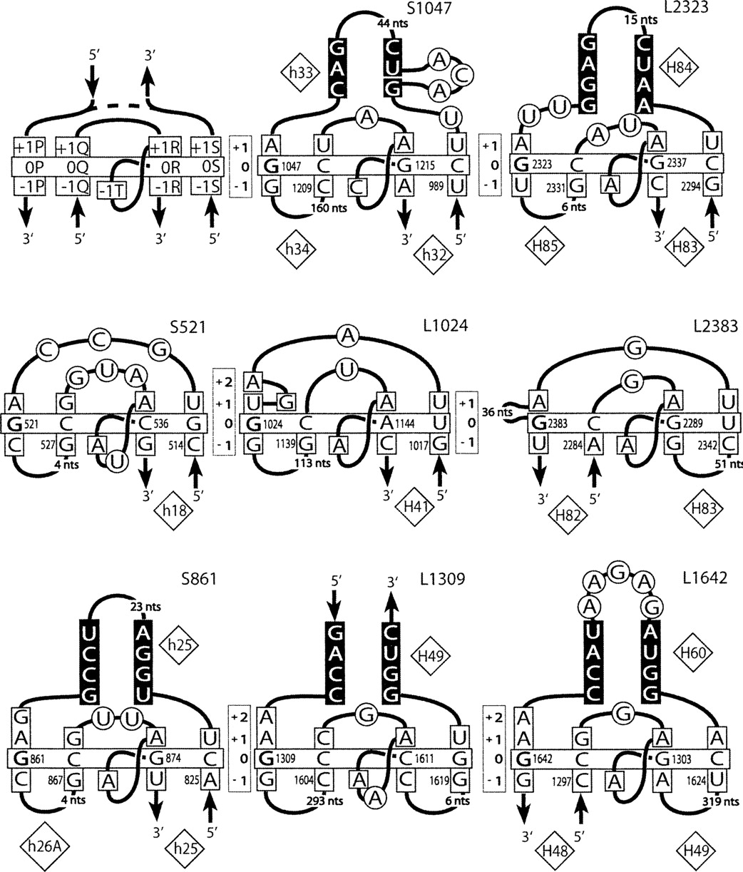
Secondary structures of the G-ribo motifs identified in the E. coli ribosome. (Upper left corner) A template with the named nucleotide positions of the layers from −1 to +1 is shown. (Dashed line) The possibility for a motif to have or not to have the third helix. In the name of a motif, “S” or “L” stands for the ribosomal subunit, small or large, in which the motif was found. The number in the name corresponds to that of nucleotide 0P in the standard E. coli numeration of rRNA. The double helices are arranged as in Figure 1A: the third helix is on top; Helices 1 and 2 are at bottom left and bottom right, respectively. (Vertically oriented rectangles) The positions of the layers. (Horizontally oriented rectangle) The base pairs at the zero layer. G in position 0P is bold. The nucleotides that stack to those of the zero layer are squared. The nucleotides of the third helix involved in base-pairing are shown on the black background. Other nucleotides are circled. The numbers of the helices in the standard 16S and 23S rRNA secondary structures are shown in diamonds.




