


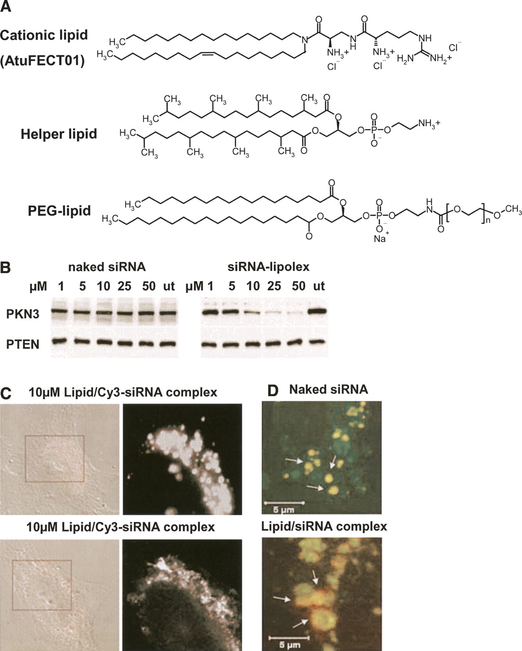
In vitro evaluation of lipid/siRNA complexes. (A) Chemical structures of lipids used for preparation of cationic liposomes and complex formation with siRNAs. (AtuFECT01) β-L-Arginyl-2,3-L-diaminopropionic acid-N-palmityl-N-oleyl-amide trihydrochoride; (helper lipid) 1,2-diphytanoyl-sn-glycero-3-phosphoethanolamine; (PEG-lipid) distearoyl-sn-glycero-3-phospho-ethanolamine sodium salt. (B) Concentration-dependent inhibition of PKN3 protein expression with lipid/siRNA complex, but not with naked siRNA in HeLa cells as assessed by immunoblot. PTEN served as the loading control. (C) Intracellular distribution of lipid/Cy3–siRNA complexes in HeLa cells analyzed by confocal microscopy. (D) Merged high-magnification pictures of a cytoplasmic area from HUVECs showing endosomal/lysosomal localization of siRNAs. Naked siRNAs remain trapped in the endosomal pathway (upper panel), whereas formulated siRNAs escape the late endosomal/lysosomal vesicles (lower panel). Reprinted by permission from Macmillan Publishers Ltd.: [Gene Therapy] (Santel et al.) © (2006).




