


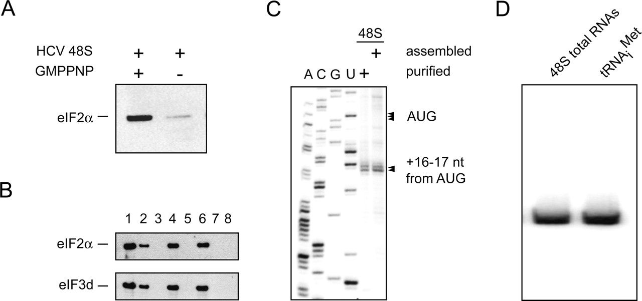
Analysis of the composition of HCV IRES 48S complexes. (A) Immunoblotting analysis of HCV IRES 48S complexes assembled in the absence and the presence of GMPPNP using anti-eIF2α antibodies. (B) Efficiency of the purification protocol. The presence of eIF2 and eIF3 is followed by immunoblotting through the entire purification. Lanes are loaded as follows: (1) application fraction, (2) flowthrough of the application fraction, (3) wash fractions, (4) elution, (5) supernatant of the centrifugation step, (6) 48S peak from the sucrose density gradient, (7) 40S/IRES peak from the gradient, (8) pooled top fractions from the gradient. (C) Toeprinting assay of both purified and assembled 48S HCV initiation complexes. Arrows denote positions of the initiation AUG codon and toeprinting stops. A dideoxynucleotide sequence generated with the same primer (shown on the left) was run in parallel. The upper part of the gel, which contains additional, previously observed toeprints (Pestova et al. 1998) caused by the processivity of the reverse transcriptase in combination with the particle stability under the assay condition (see Materials and Methods), is omitted for clarity. (D) Detection of Met-tRNAiMet within initiation complexes. Northern blot analysis of HCV IRES 48S compared with a transcribed tRNAiMet.




