


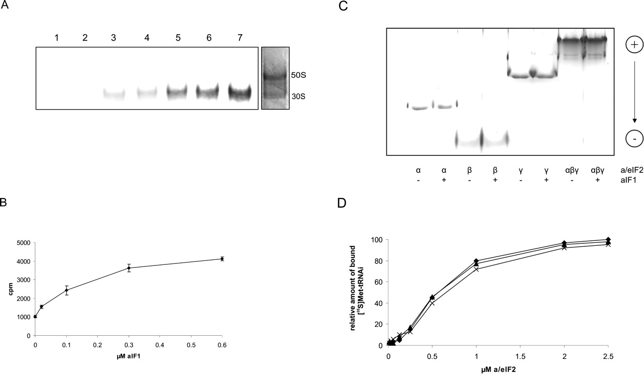
NH-aIF1 stimulates Met-tRNAi binding to ribosomes but not to purified a/eIF2. (A) S. solfataricus ribosomes (0.1 μM) were incubated with a/eIF2·GTP· [35S]Met-tRNAi (lanes 3–7) and increasing amounts of aIF1 (0.02, 0.1, 0.3 and 0.6 μM, respectively) (lanes 4–7) at 70°C. (Lane 1) [35S]Met-tRNAi (1 μM) was incubated with ribosomes (0.1 μM). (Lane 2) aIF1 (1 μM) was incubated together with [35S]Met-tRNAi (1 μM) and ribosomes (0.1 μM). Samples were loaded on a native 4% polyacrylamide gel, and the radioactive bands were visualized using a PhosphorImager. One representative experiment is shown. The positions of the 30S and 50S ribosomal subunits in the gel were revealed in a parallel experiment by Coomassie blue staining. The positions of the 50S and 30S ribosomal subunits are indicated. (B) Graphical representation of two independent experiments of [35S]Met-tRNAi binding to ribosomes as a function of the aIF1 concentration. The error bars represent standard deviations. (C) The α-, β-, and γ-polypeptides or reconstituted a/eIF2 trimer (50 pmol each) were incubated at 65°C for 15 min in the presence (+) or absence (−) F1 (100 pmol). The samples were electrophoresed on a nondenaturing polyacylamide gel as described in Materials and Methods. The pI of aIF1 is ~7; therefore, free protein does not enter the gel per se. Migration of the proteins from the cathode toward the anode is indicated on the right. (D) Increasing amounts of S. solfataricus a/eIF2 were incubated with GTP (1 mM) and [35S]Met-tRNAi (1μM) at 70°C in the absence of aIF1 (diamonds) or in the presence of 2 (triangles) and 5 (crosses) μM aIF1, respectively. The amount of [35S]Met-tRNAi bound by 2.5 μM a/eIF2 in the absence of aIF1 was set to 100%.




