


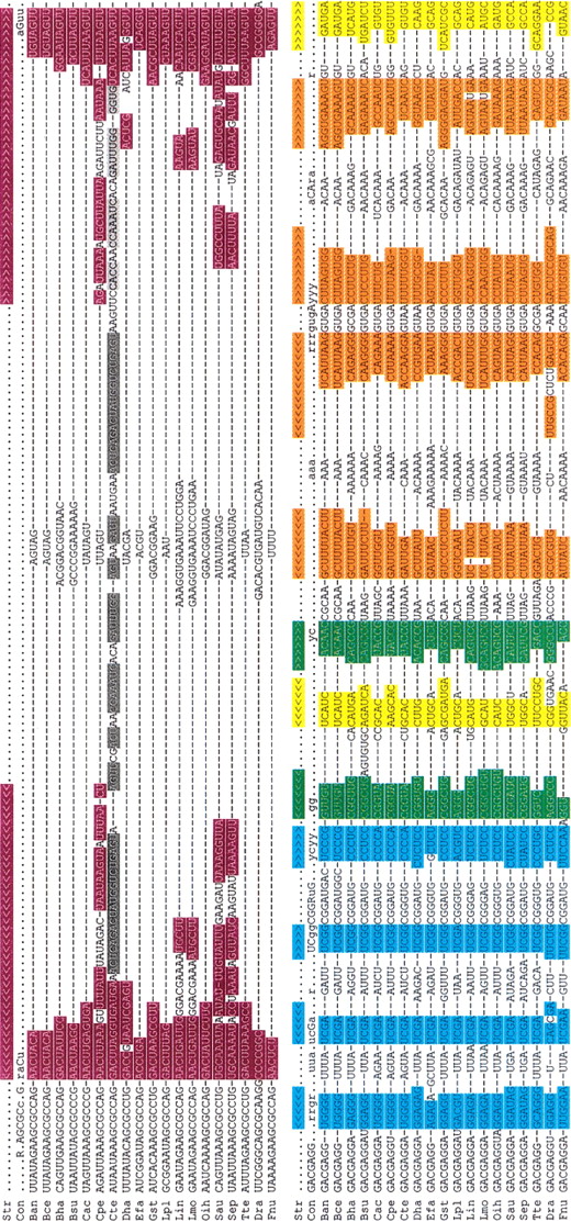
Nucleotide sequences and alignment of glmS ribozymes from 19 bacterial species (expanded from data in Barrick et al. 2004). The 5′ and 3′ portions of each conserved secondary structure element are color-coded as follows: P1, violet (P1 extensions are highlighted in gray); P2, blue; P3, green; P3a (pseudoknot), yellow; P4, orange. The locations of complementary nucleotides are indicated with braces in the structure line (Str). The consensus row (Con) designates the identities of nucleotides that are 100% conserved (uppercase) and >75% conserved (lowercase) among the aligned sequences (R = A, G; Y = C, U). Organism abbreviations are as follows: (Ban) Bacillus anthracis, (Bce) Bacillus cereus, (Bha) Bacillus halodurans, (Bsu) Bacillus subtilis, (Cac) Clostridium acetobutylicum, (Cpe) Clostridium perfringens, (Cte) Clostridium tetani, (Dha) Desulfitobacterium hafniense, (Efa) Enterococcus faecalis, (Gst) Geobacillus stearothermophilus, (Lpl) Lactobacillus plantarum, (Lin) Listeria innocua, (Lmo) Listeria monocytogenes, (Oih) Oceanobacillus iheyensis, (Sau) Staphylococcus aureus, (Sep) Staphylococcus epidermidis, (Tte) Thermo-anaerobacter tengcongensis, (Dra) Deinococcus radiodurans, (Fnu) Fusobacterium nucleatum.




