


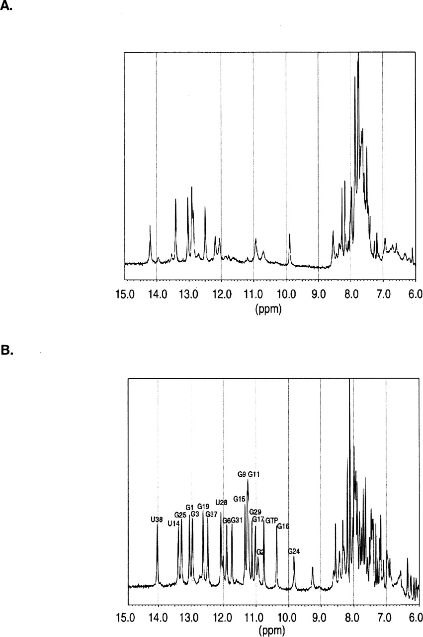
FIGURE 8.
1D exchangeable and aromatic proton spectra for the Class I GTP aptamer. (A) Spectrum collected before GTP was added to the sample. (B) Spectrum collected after a 1.1-fold molar excess of GTP was added to the sample. The spectra were collected at 500 MHz in 95% H2O/5% D2O at 24°C. Residues with well-resolved imino proton resonances are identified. (See also Fig. 2 of Dieckmann et al. 1996.)




