


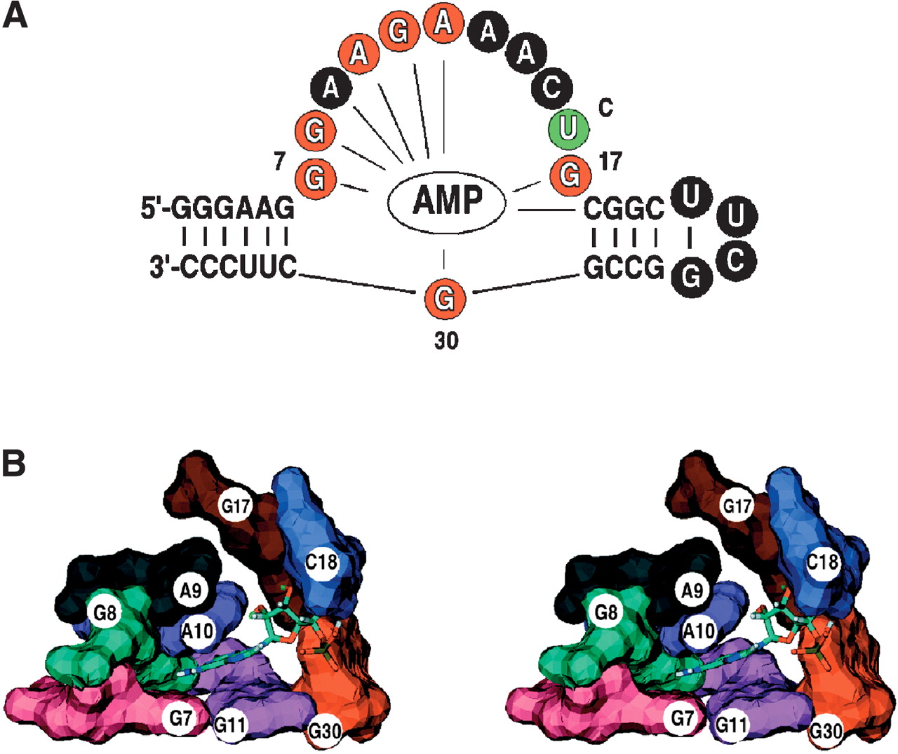
FIGURE 7.
Sassanfar ATP aptamer (Sassanfar and Szostak 1993). (A) The secondary structure model is shown in the same manner as the Class I GTP aptamer in Figure 1. The recognition bulge nucleotides are colored according to sequence conservation. The lines indicate that intermolecular NOEs to the AMP ligand were detected for those positions (Dieckmann et al. 1996). (B) Stereo view of the molecular surface of the Sassanfar ATP aptamer binding pocket (PDB ID 1RAW) with the AMP ligand CPK colored and residues numbered as in A.




