


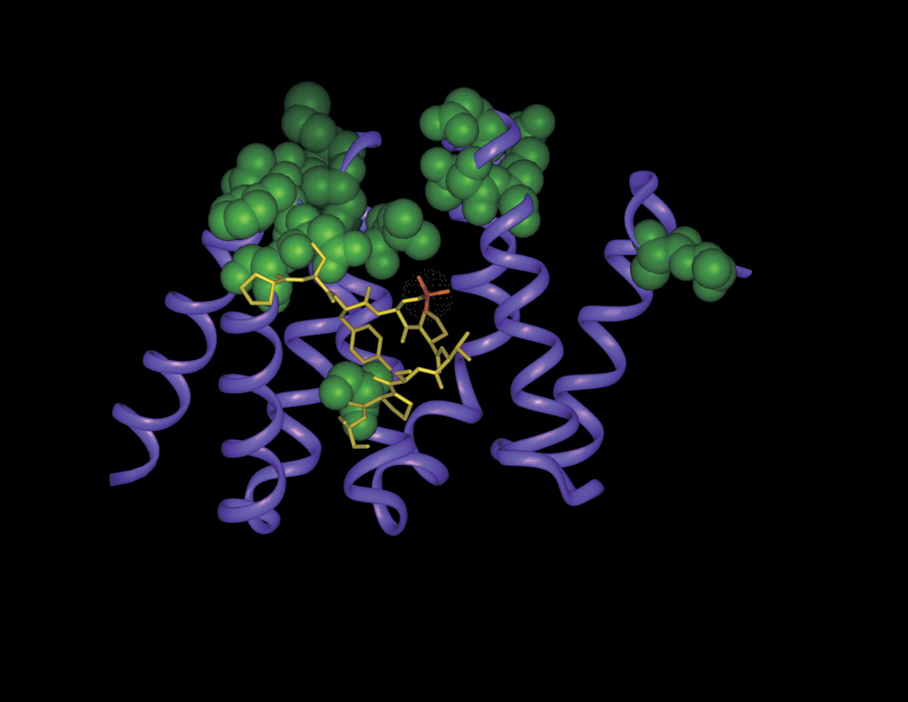
FIGURE 2.
Ribbon representation of Pcf11 CID (magenta) in complex with the CTD-derived peptide (Meinhart and Cramer 2004). The peptide residues are shown in yellow and phosphoserine 2 is highlighted in orange. The CID residues whose resonances are most affected during the titration with GA-RNA are displayed using a CPK render (green). The binding sites of the CTD-peptide and of the RNA partially overlap.




