


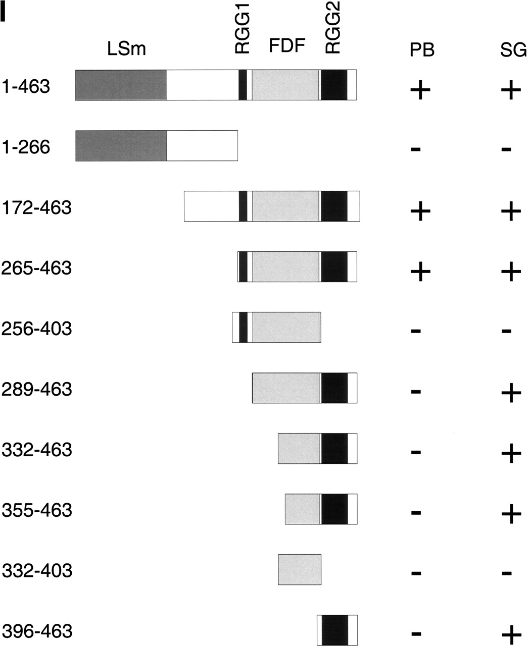
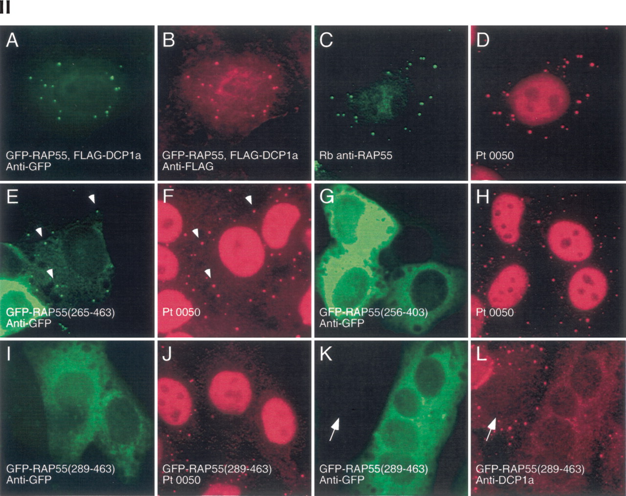
Structure of RAP55 and identification of the portions of RAP55 that mediate localization to P-bodies and stress granules. (Panel I) The predicted amino acid sequence of RAP55 contains an N-terminal LSm domain and two C-terminal RGG-rich domains. Between the two RGG domains is an FDF motif. The portions of RAP55 that were capable of localizing green fluorescent protein (GFP) to P-bodies (PB) or stress granules (SG) are indicated by +. Representative indirect immunofluorescence results are shown in panel II. (Panel II) Two-color immunofluorescence was used to identify the portions of RAP55 that were necessary and sufficient for localization to P-bodies. (A) Full-length GFP-RAP55 localized to cytoplasmic dots and (B) colocalized with cotransfected DCP1a. (C) Rabbit anti-RAP55 antibodies colocalized with Ge-1-containing P-bodies as determined (D) using human serum 0050, which reacts with Ge-1, but not RAP55. A C-terminal portion of RAP55 (amino acids 265–463) (E) localized to P-bodies and (F) colocalized with Ge-1. White arrows indicate examples of overlapping cytoplasmic foci. (G) RAP55(256–403), which lacks the RGG2 domain, was present diffusely throughout the cytoplasm and did not localize to P-bodies. (H) RAP55(256–403) did not alter the P-body location of Ge-1. (I) RAP55(289–463) also did not localize to P-bodies, but this portion of RAP55 (J) displaced Ge-1 from P-bodies. Similarly, (K) RAP55(289–463) displaced DCP1a from (L) P-bodies in transfected cells. Note that DCP1a-containing P-bodies were detected in nontransfected cells (indicated by white arrows in K and L). Successful production of the GFP-RAP55 fragment fusion proteins used in these studies was confirmed by immunoblot using anti-GFP antiserum (data not shown).




