


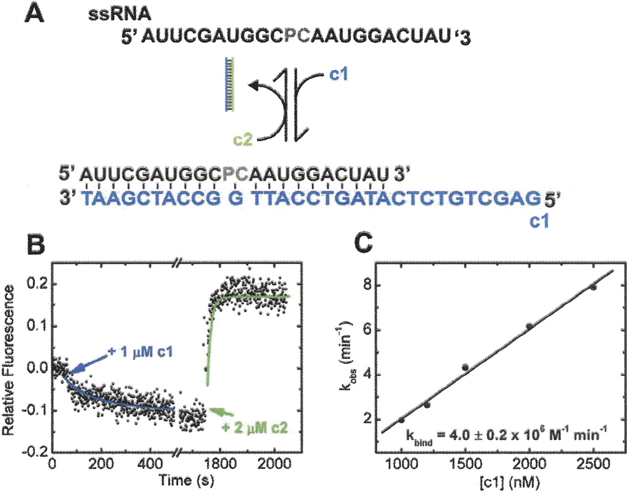
Kinetics of formation and dissociation of an RNA/DNA duplex in standard buffer (50 mM Tris, 25 mM MES-NaOH, 25 mM acetic acid–NaOH at pH 7.5, 100 mM NaCl) at 25°C. This assay is analogous to previous conformational shift assays (Yurke et al. 2000; Hoerter et al. 2004) and demonstrates the reversibility of changes in pyrrolo-C fluorescence upon duplex formation. (A) Schematic of the assay. ssRNA binds DNA oligonucleotide c1 (cyan), which is removed by addition of DNA oligonucleotide c2 (green); gray indicates pyrrolo-C probe. (B) Upon addition of the DNA oligonucleotide c1 (cyan), the pyrrolo-C fluorescence decreases with a rate of 1.93 min−1. Upon addition of DNA oligonucleotide c2 (green), the fluorescence decrease is reversed, with a rate of 4.28 min−1. (C) Concentration dependence of the pseudo-first-order rate constant upon addition of excess c1 to ssRNA. The slope of the linear regression line yields the bimolecular rate constant kbind.




