


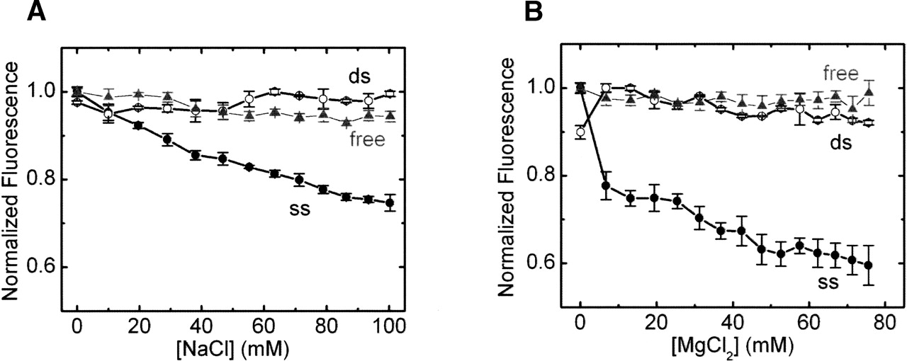
FIGURE 3.
Relative fluorescence intensity of free pyrrolo-C ribonucleoside (gray triangles), ssRNA (filled circles), and dsRNA (open circles) as a function of NaCl (A) and MgCl2 (B) concentration in 50 mM Tris, 25 mM MES-NaOH, and 25 mM acetic acid–NaOH (pH 7.5; initially containing ~12 mM Na+) at 25°C. Errors are derived from at least two independent measurements.




