


FIGURE 2.
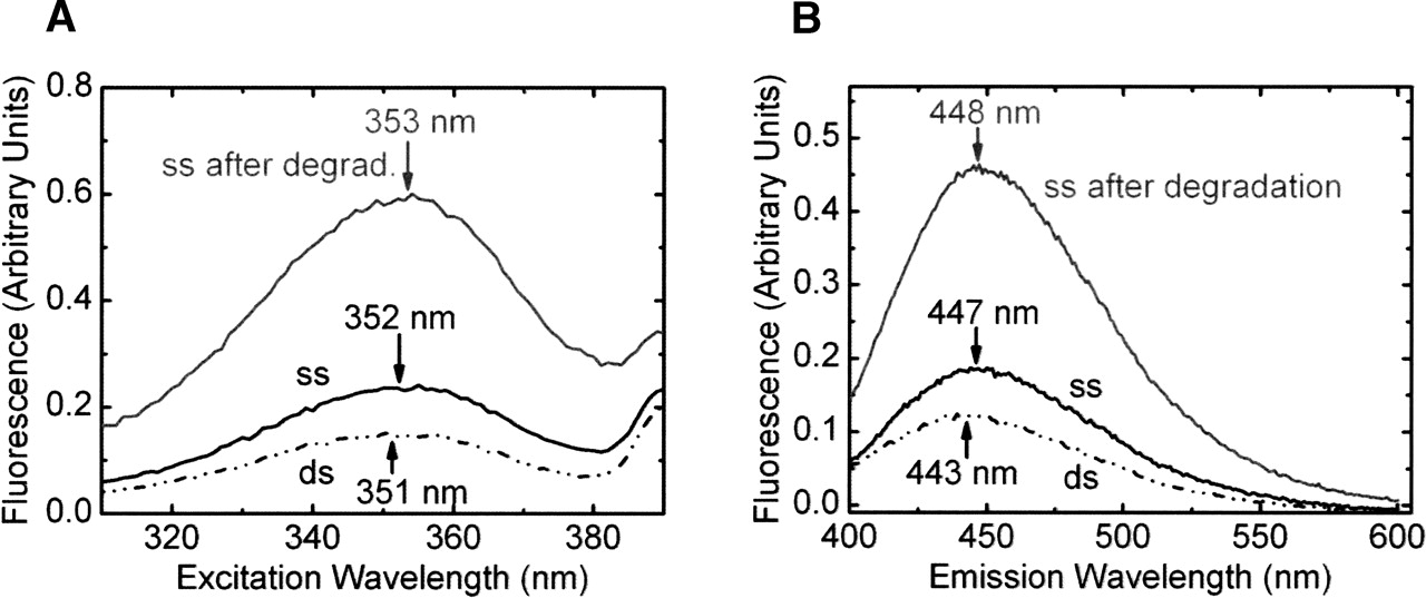
Pyrrolo-C fluorescence studies using 1 μM ssRNA and 1 μM dsRNA in standard buffer (50 mM Tris, 25 mM MES-NaOH, 25 mM acetic acid–NaOH at pH 7.5, 100 mM NaCl) at 25°C. Steady-state fluorescence emission (A) and excitation spectra (B) of ssRNA (solid line), dsRNA (dash-dotted line), and ssRNA after complete degradation with 0.5 U of S1 nuclease (gray line), as indicated. The emission and the excitation maxima were obtained as the means of Gaussian fits to the spectra.




