


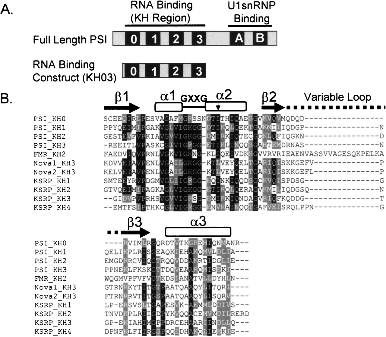
FIGURE 2.
PSI construct and sequence info. (A) A comparison of the full-length PSI protein and the construct used for binding experiments in this study. (B) Alignment of KH domains from several proteins showing conserved residues and secondary structure motifs. Arrow denotes location of conserved Ile residue mutated to Asn in individual PSI KH domains. Secondary structure was based on that observed by Lewis et al. (2000).




