


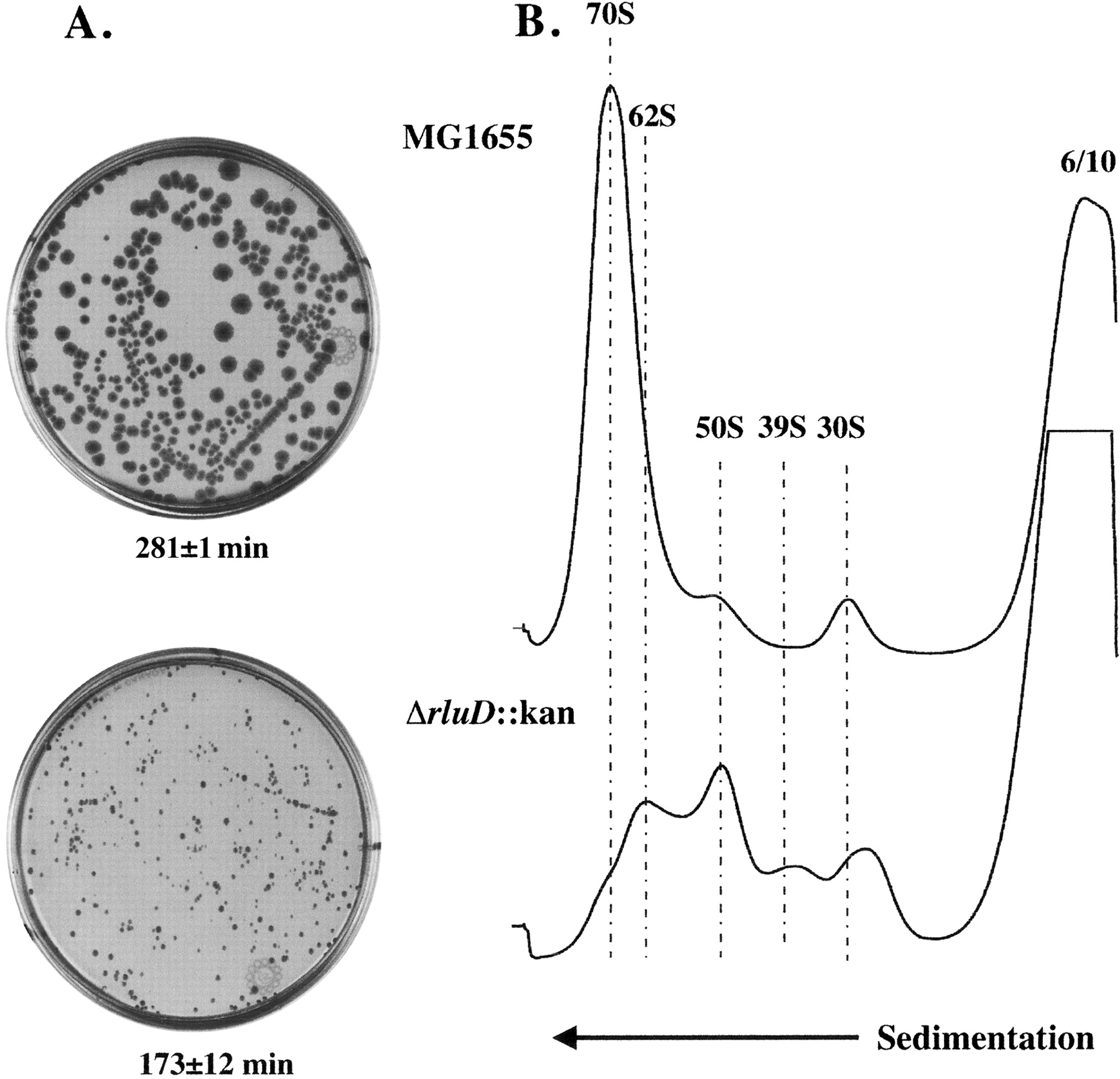
Colony morphology, exponential growth rates and ribosome profiles of wild-type and ΔrluD::kan mutant E. coli strains. (A) LB plates without drug (wild type) or with 34 μg/mL kanamycin (mutant only) were incubated 41 h at 37°C. Dilutions were prepared from suspensions of wild-type and mutant cells and 6.8 × 10⁸ cells/mL for each strain were spread on plates and incubated. Exponential growth rates were measured by monitoring cell density at 600 nm with a Perkin Elmer Lambda 25 UV/VIS Spectrophotometer. Doubling time was determined from a semilogarithmic plot of A600 versus time. Each plot consisted of five to seven time points. Doubling times were the average of two determinations (wild type) or six determinations (mutant), and are shown under the plates. (B) Ribosome profiles of wild-type and ΔrluD::kan cells were performed as described in Materials and Methods. Wild-type and mutant cells were lysed at 6 mM Mg++ and gradients were run at 10 mM Mg++. Twelve A260 units were layered onto 14%–32% sucrose gradients and centrifuged at 22,000 rpm for 19 h at 4°C.




