


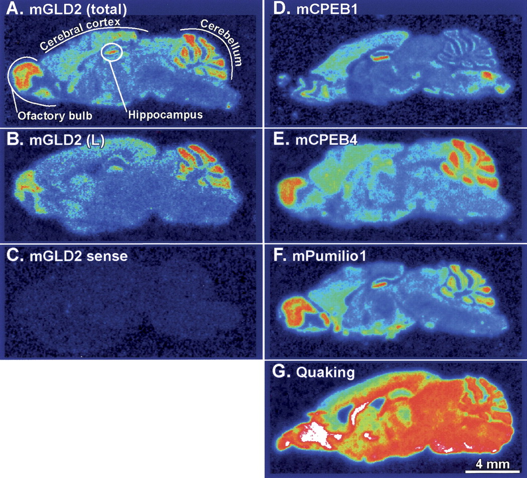
The pattern of mGLD2 mRNA abundance in neuronal cell populations in the mouse brain is similar to those of mCPEB1 and mPumilio1 mRNAs. (A) In situ hybridization on sagittal mouse brain sections using an 35S-labeled mGLD2 RNA probe that detects total mGLD2 mRNA (i.e., mGLD2(L) plus mGLD2(S)). The detected pattern of mGLD2 mRNA is consistent with expression in the major neuronal cell layers in the olfactory bulb, cerebral cortex, cerebellum, and hippocampus, each of which is labeled. (A–G) Bar, 4 mm. (B) Same as A, but using a probe specific to the unique region of the 3′UTR of mGLD2(L). (C) Same as A, but hybridized to a probe with the same sense as mGLD2 mRNA. (D–G) Adjacent sections hybridized with the indicated probes to detect mCPEB1 (D), mCPEB4 (E); Pumilio1 (F) and Quaking (G) mRNAs.




