


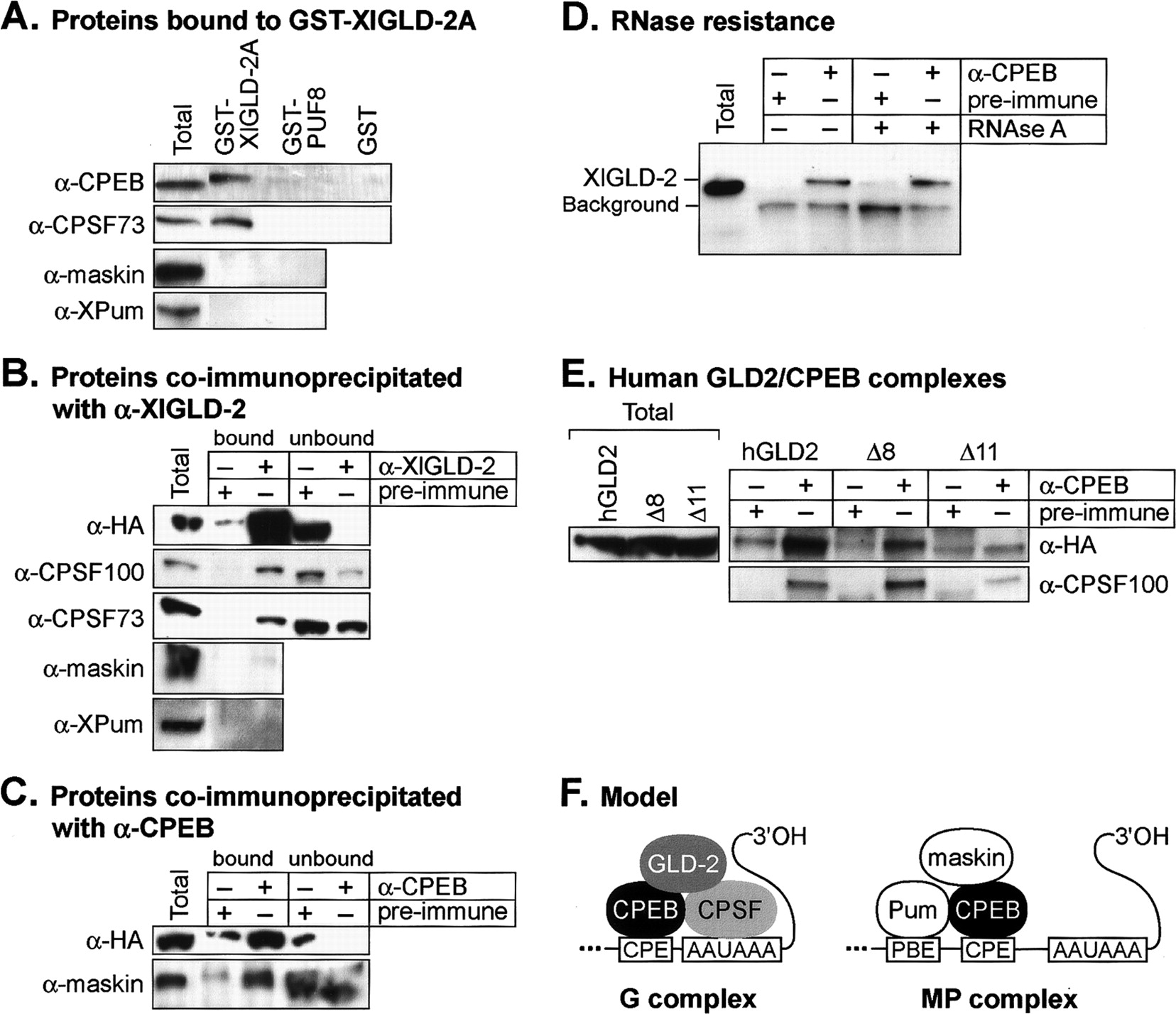
XlGLD-2 interacts with polyadenylation factors. (A) Proteins bound to GST-XlGLD-2A. Stage VI oocyte lysate was inubated with purified GST-XlGLD-2A, GST-PUF-8, or GST, bound to glutathione-Sepharose. Proteins retained on beads after three washes were analyzed by Western blotting with antibodies specific to CPEB, CPSF-73, maskin, and XlPu-milio. Total indicates unfractionated lysate (fourfold less starting material than in the other lanes). (B) Proteins coimmunoprecipitated with α-XlGLD-2. Crude lysates were prepared from oocytes that were expressing HA/XlGLD-2. Lysates were incubated with α-XlGLD-2 (top panel) or α-CPEB (bottom panel), or with respective preimmune sera, as indicated above each lane. Proteins were detected by Western blot analysis using α-CPSF100, α-CPSF73, α-maskin, and α-XlPumilio, as indicated. Western analysis using α-HA as probe demonstrated that the α-XlGLD-2 antibody efficiently immunoprecipitates the endogenous protein. Bound indicates proteins that bound to the beads; unbound, proteins that did not bind to the beads. Total indicates unfractionated lysate (threefold less starting material than in bound, and equal starting material to unbound). (C) Proteins coimmunoprecipitated with α-CPEB. Oocytes were injected with mRNAs that encode HA-XlGLD-2. Oocyte lysates were incubated with α-CPEB, or with preimmune serum. Both XlGLD-2 and maskin are associated with α-CPEB. Total indicates unfractionated lysate (threefold less starting material than in bound, and equal starting material to unbound). (D) Ribonuclease-resistance of the CPEB/GLD-2 interaction. Xenopus oocyte extracts, prepared as in B, were treated with RNAseA. Gel electrophoresis demonstrated that the RNA had been degraded (data not shown). Coimmunoprecipitations were performed with α-CPEB or preimmune sera, on X. laevis oocyte extracts as in B. The two proteins continue to coimmunoprecipitate after RNAse treatment. (E) Human GLD2/CPEB complexes. Immunoprecipitations using either α-CPEB or preimmune antibodies were incubated with X. laevis oocyte extracts prepared from oocytes expressing either HA-hGLD2, HA-hGLD2Δ8, or HA-hGLD2Δ11. Total indicates crude lysate. (F) Model of polyadenylation complexes in resting oocytes. See text for details. CPSF and CPEB are shown interacting in the resting oocyte (Mendez et al. 2000; Dickson et al. 2001). However, CPEB phosphorylation increases binding affinity to CPSF (Mendez et al. 2000). Symplekin is likely to be in the G complex, as inferred from the data of Barnard et al. (2004).




