


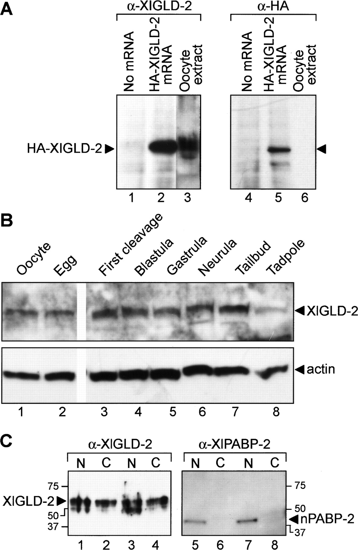
XlGLD-2 is present throughout embryogenesis and is in the cytoplasm. (A) Antibody specificity. mRNA encoding HA-XlGLD-2A was translated in vitro, and the products were analyzed by Western blotting using α-XlGLD-2 antibodies. A predominant protein of 60 kDa is detected by α-XlGLD-2 (lanes 1–3) and α-HA antibodies (lanes 4–6). The in vitro translated protein (lane 2) roughly comigrates with endogenous XlGLD-2 protein in oocyte extracts (lane 3). (B) XlGLD-2 protein abundance. Proteins extracted from the indicated stages were analyzed by Western blotting using either α-XlGLD-2 or α-actin. (C) XlGLD-2 subcellular distribution. Nuclear and cytoplasmic fractions prepared manually from stage VI oocytes were analyzed by Western blotting with α-XlGLD-2 (lanes 1–4). α-XlPABP-2 (lanes 5–8), which detects a nuclear poly(A) binding protein (Good et al. 2004), was used to control for proper enucleation and a lack of leakage into the cytoplasm. N and C indicate nucleus and cytoplasm, respectively.




