


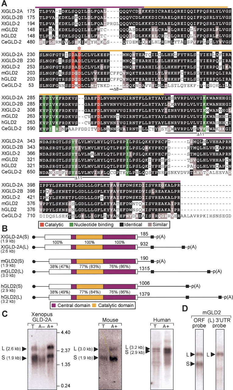
XlGLD-2, mGLD2, and hGLD2 mRNAs and proteins. (A) Predicted protein sequences in the catalytic and central domains of GLD2 homologs. The sequences from five animal different species (X. laevis, X. tropicalis, mouse, human, and C. elegans) are presented. Black indicates amino acid identity; red, three carboxylate side-chains required for catalysis; green, six residues that position the nucleotide (Martin et al. 2000; Wang et al. 2002); and red asterisk, location of the active site mutation, D242A. Colored bars above the sequence indicate the central (purple) or catalytic (yellow) domains. Black lines below the sequence indicate the regions missing in Δ8 and Δ11 forms of the human protein. (B) Vertebrate GLD2 mRNAs. Approximate lengths of mRNAs, as calculated by cDNA sequencing and confirmed by Northern blotting, are shown to the left of each panel. Purple indicates PAP central domain; yellow, PAP catalytic domail (corresponding to Fig 1A). Percentage of amino acid sequence identity relative to XlGLD-2A is given (percentage similarity is in parentheses). Black circles and boxes indicate 5′ cap and 3′ cleavage and polyadenylation signal (AAUAAA and AAUACA). Distances from the termination codon to the poly(A) tail are indicated. (C) Two mRNA forms. RNAs from Xenopus oocytes, mouse 3T3 cells, and human spleen were analyzed by Northern blotting. T indicates total RNA; A+, RNA retained by oligo(dT) cellulose; and A-, RNA not retained by oligo (dT). Amounts of RNA and hybidization probes are described in Materials and Methods. (D) mGLD2(L) and mGLD2(S) mRNAs differ by their 3′UTRs. Total RNA from spleen was analyzed by using either a probe complementary to the entire ORF (left) or just the 3′UTR of the long form of mRNA, mGLD2(L) (right).




