


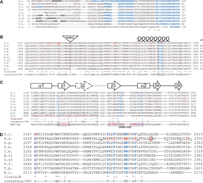
Alignments of 10 Prp8 orthologs showing four conserved motifs. Important residues are highlighted in bold blue type and known alleles underlined in red. (S.c.) Saccharomyces cerevisiae; (H.s.) Homo sapiens; (P.f.) Plasmodium falciparum; (D.m.) Drosophila melonagaster; (C.e.) Caenorhabditis elegans; (A.t1.) Arabidopsis thaliana chromosome 1; (O.s5) Oryza sativa chromosome 5; (S.p.) Schizosaccharomyces pombe; (T.b.) Trypanosoma buceii; (E.c.) Encephalitazoon cuniculii. Numbers refer to the amino acid residues. All sequences aligned using ClustalW and consensii are derived from 25 Prp8 orthologs using the ‘consensus’ program. (A) Alignment of the N-terminal regions. The polyproline tracts can be seen in the nonconserved N termini of some species. Potential bipartite NLS sequences are highlighted in bold blue type and are separated by 15 amino acids. In the case of S. cerevisiae an overlapping nuclear localization signal (NLS) sequence is underlined. In the cases of P. falciparum and E. cuniculi, a shorter SV40 type NLS and pat7 types are highlighted, respectively. The NLS sequences always occur within the first 500 N-terminal residues as predicted by PSORTII (Nakai and Horton 1999). (B) The highly conserved Prp8 domain 3.2. The position of the intein, present in four orthologs is indicated by the triangle. A potential protein kinase C phosphorylation site (T/S-x-R/K) that is conserved in all species is boxed and highlighted. This is central to the postulated coiled-coil domain represented above the text by the coil. (y%) The level of conservation compared with the yeast sequence. (C) The MPN/JAB domain found in Prp8. The mutations yprp8–28 and sprp8spp42–1 are shown in bold red underlined text. The representative MPN/JAB domain from NCBI’s domain database is shown at the bottom of the alignment. Two important MPN motif residues identified previously are boxed in plum coloured text (Maytal-Kivity et al. 2002). The five residues in the MPN structure that coordinate the zinc are highlighted in bold blue text. The secondary structure diagrams are shown above the alignment (from Tran et al. 2003). (D) The C terminus of Prp8 showing the single mutated residues altered in human Retinitis Pigmentosa (bold, red underlined type), the nonsense mutation (bold, red, and circled) and the residues where frameshift mutations occur (doublly underlined text). The temperature-sensitive yprp8–1 mutation affects the first glycine residue on the left-hand side of the alignment (bold plum).




